
General Mechanism of Actions of INSULIN.

GENERAL MECHANISM OF ACTION OF INSULIN AND RELATE HORMONES

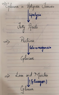
  • The body's main fuel is Glucose. Glucose is stored in the liver and muscles as Glycogen. 
  • Excess glucose is stored in the adipose tissues which yield fatty acids via lipolysis. In the fasting state, free fatty acids supply much of the energy needs of the body except for the CNS which requires Glucose to function normally.
  • Proteins can also be converted to glucose via gluconeogenesis.

RELEASE OF INSULIN AND GLUCAGON

  • Insulin and Glucagon are produced in the pancreas by the Islets of Langerhans. Beta cells make up 70%-90% of the Islets and produce Insulin and Amylin while the a-cells produce glucagon.
  • The main function of insulin is to decrease blood glucose levels.
  • Glucagon, along with other counter-regulatory hormones such as growth factor, cortisol, and Epinephrine, increases Blood glucose levels. 
  • The opposing actions of Glucagon and Insulin along with other counter-regulatory hormones maintain a normal fasting values of 79-99mg/dL.

FUNCTION OF INSULIN/ NORMAL INSULIN ACTION

After the food is consumed, blood glucose levels rise, and the secretion of insulin occurs in two phases:

Phase 1-FIRST PHASE INSULIN RESPONSE:

An initial burst lasts approx. 5-10 minutes and serves to suppress hepatic glucose production and causes insulin-mediated glucose disposal in adipose tissues. This bolus of insulin minimizes hyperglycemia during meals and during post-prandial period.

Phase 2: 

The second phase is characterized by the gradual increase in insulin secretion, which lasts 60-120 minutes and stimulates the glucose uptake by Peripheral insulin dependent tissues. 


FUNCTION OF AMYLIN

  • Amylin is the naturally occurring hormone that is co-secreted by the beta cells along with Insulin. 
  • People with diabetes either have a relative or complete lack of amylin.
  • 3 major functions of Amylin are: 
  • a) Suppression of post meal glucagon secretion 
  • b) Regulation of the rate of gastric emptying from the stomach to the small intestine, which increases satiety.
  •  c)Regulation of plasma glucose concentrations in the bloodstream.


Comments

Popular posts from this blog

Diabetes Mellitus: Types!

TYPES OF DIABETES MELLITUS 1. TYPE1 DM Type 1 DM is usually diagnosed before age 30 years, but it can develop at any age. T1DM is an autoimmune disease in which insulin producing B-cells are destroyed. The presence of Human Leucocyte Antigens (HLAs ) is associated with the development of T1DM. In addition, these individuals often develop Islet cell antibodies , Insulin Autoantibodies or Glutamic Acid decarboxylase autoantibodies . At the time of development of diagnosis of T1DM, it is usually believed that most of the patients have 80-90%loss of beta cells. The remaining beta cells function at the diagnosis creates a " HONEYMOON PERIOD" during which the blood glucose levels are easier to control, and smaller amounts of insulin are required. after this, the remaining beta cells function is completely lost, and the patients become completely deficient of insulin and hence require exogenous insulin.  2. TYPE1.5 DM:  Also referred as Latent autoimmune diabetes in Adults (LAD) ...

Diabetes Mellitus: Pharmacotherapy- Gliptins and a-Glycoside Inhibitors!

  PHARMACOTHERAPY:  Medications classifications include:  Sulfonyl Urea's Nonsylfonylurea Secretagogues (a-Glinides) Biguanides Thiazolidinediones a-Glucosidase Inhibitors Dipeptidyl Peptidase-4 Inhibitors (Gliptins) Sodium-glucose co-transporter (SGLT) Inhibitors  Central-Acting Dopamine Agonists Bile Acid Sequestrants Insulin therapy 5. a-GLUCOSIDASE INHIBITORS Acarbose and Miglitol are a-glucosidase inhibitors currently approved in the United States. Mechanism of Action: An enzyme that is along the brush border of the intestine cells called a-glucosidase; breaks down the complex carbohydrates into simple sugars, resulting in absorption. The a-glucosidase inhibitors work by delaying the absorption of carbohydrates from the intestinal tract, which reduces the rise in postprandial glucose levels. Therapy: As a monotherapy , a-glucosidase inhibitors primarily reduce post-prandial glucose excursions.  Clinical Use: FPG concentrations have been decreased by 40-5...

Drug-Induced Nephrotoxicity!

 DRUG-INDUCED NEPHROTOXICITY Drug-Induced Nephrotoxicity is increasingly recognized as a significant contributor to kidney disease including Acute Kidney Injury (KI) and chronic kidney disease (CKD). Nephrotoxicity has a wide spectrum, reflecting damage to different nephron segments based upon individual drug-mechanisms. 1. ACE-INHIBITORS: ACE-Inhibitors are frequently associated with Proteinuria and Renal Insufficiency. The prevalence of Proteinuria in Captopril treated patient is estimated to be 1%. The risk of Renal Insufficiency is greater with long-acting ACE Inhibitors such as Lisinopril or Enalapril than with Captopril. Immune complex glomerulopathy is a major contribute to ACE Inhibitor nephrotoxicity. Predisposing factors include: Hyponatremia Diuretic therapy Pre-existing renal impairment Congestive Heart Failure (CHF) Diabetes Mellitus Recovery of renal function usually follows ACE-Inhibitor discontinuation. 2. CEPHLOSPORIN: The cephalosporin antibiotics are capable of...